
Dr. Srino Bharam is a board-certified fellowship-trained sports medicine orthopedic surgeon specializing in the treatment of athletic injuries of the hip for teens and adults. Dr. Bharam is a pioneer of hip arthroscopy with over 30 years of experience in treating injuries and conditions of the hip with the goal of restoring athlete and patients to an active lifestyle.
Your hip is built to take a lot of wear-and-tear, it’s a very complex joint. Athletes and other active people put a lot of stress on the joint and pain in the hip or groin region can result. Gradual damage is also common as we age and pain in this area can be severe. Because this pain can come from many sources and can be felt in many parts of the hip region, it’s important to understand the underlying conditions that can cause it.
As Dr. Bharam has noted, hip injuries are often misdiagnosed. He notes that one study saying that patients are typically seen by more than three different health professionals before a torn labrum is correctly diagnosed. Aside from the frustration of an incorrect diagnosis, additional damage can occur in the meantime. That’s why it’s important to start with the level of expertise found at the Hip & Groin Preservation Center.
Anatomy
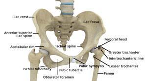
The thigh bone, femur, and the pelvis, acetabulum, join to form the hip joint. The hip joint is a “ball and socket” joint. The “ball” is the head of the femur, or thigh bone, and the “socket” is the cup shaped acetabulum. The joint surface is covered by a smooth articular surface that allows pain free movement in the joint.
Cartilage Acetabulum “The Socket”
The cartilage cushions the joint and allows the bones to move on each other with smooth movements. This cartilage does not show up on X-ray, therefore you can see a “joint space” between the femoral head and acetabular socket.
Pelvis
The pelvis is a large, flattened, irregularly shaped bone, constricted in the center and expanded above and below. It consists of three parts: the ilium, ischium, and pubis.
The socket, acetabulum, is situated on the outer surface of the bone and joins to the head of the femur to form the hip joint.
Femur
The femur is the longest bone in the skeleton. It joins to the pelvis, acetabulum, to form the hip joint.
The upper part is composed of the femoral head, femoral neck, and greater and lesser trochanters.
Labrum
The acetabular labrum is an elastic cartilage that lines the edge of the acetabulum (socket), providing stability to the joint.
The labrum deepens the socket, holds the joint fluid inside the socket and forms a protective cushion between the acetabulum and the constantly moving femoral head.